TREATED DISEASES
⦁ Low back pain and low back pain
⦁ Cervicalgia and cervicobrachialgia
⦁ Stenosis of the vertebral canal
⦁ Ernie or disc protrusions
⦁ Vertebral fractures
⦁ Joint veneer syndrome
⦁ Inflammation of the sacroiliac joint
⦁ Headache
TREATMENTS:
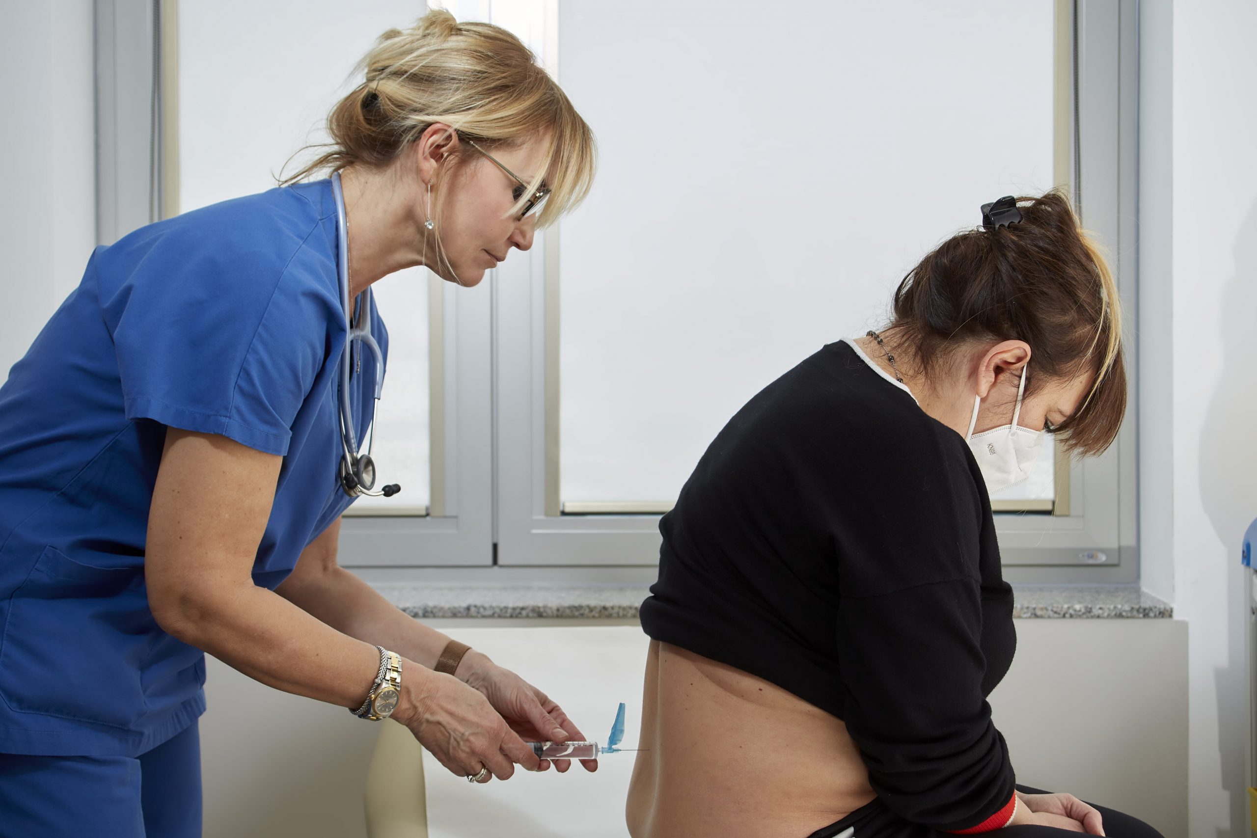
⦁ Infiltrative therapies
⦁ Acupuncture
⦁ Denervation of joint veneers
⦁ Peridural
⦁ Perhydroscopy
⦁ Intradiscal treatments
⦁ Cryotherapy
⦁ Denervation of the sacroiliac articulation
PAIN PROBLEMS
Back pain is one of the most widespread problems in the world population, 75-85% of Italians have suffered or suffer from some form of back pain during their life.
Back pain is the second reason for requesting a medical examination and, after the flu, it is the main cause of loss of working days in people under 45 years of age.
It affects the lumbar and sacral region; if it radiates to the lower limb it is commonly referred to as lombosciatalgia.
Pain that originates from the lumbar region can be produced by several causes, excessive exertion or a fall that produces trauma; or chronic degenerative diseases (e.g. osteoarthritis or discopathies).
The body’s response to pain is often associated with a contracture of the lumbar muscles, which increases the painful sensation, accompanying it with a marked difficulty in the movement of the trunk.
Pain is commonly divided into:
– acute, when it lasts 2/3 months and at the end of treatment it fully heals quickly
– if it persists after 3 months and must be treated with specific remedies
Most episodes of acute lumbar pain are due to excessive stretching of muscles or ligaments.
It is a condition due to the gradual narrowing of the vertebral canal,which occurs asa consequence of the degeneration of both intervertebral discs and joint veneers.
Bone spurs are formed, called osteophytes,as a result of excessive loadon the intervertebral disc and for the progress of osteoarthritis of the spine (spondylosis).
These osteophytes tend to reduce the diameter of the vertebral canal.
The joint veneers widen and deform, further reducing the space available to the nerve roots and spinal cord.
As stenosis progresses, this process can lead to contact and compression of nerve roots and spinal cord.
The reason why this causes pain and weakness in the legs is still not known exactly, it can be associated with compression of the vascular vessels that carry blood to the nerve roots, which innervated the muscles.
It is a pathology of the intervertebral disc in which the inner part, called the “pulpy nucleus “, comes out of its seat makingits way through the lamellae of the fibrous anulus. Theanulus is the structure responsible for containing the nucleus and constitutes the outer contour of the disk.
If the spillage of the nucleus compresses a nerveroot, the pain can spread into the territory innervated by that root. In fact, between two vertebrae there is always a pair of spinal nerves that then go to innervate a specific area of the body.
In turn, compression of one of these spinal nerves causes pain in the part of the body that they innervate.
The two positions in which the vast majority occur (about 98%) of the painful disc hernias are the last two intervertebral spaces, namely L4-L5 and L5-S1.
Most episodes of discanulus rupture occur between the age of 30 and 40, when the consistency of the nucleus is still essentially gelatinous.
The most common type of vertebral fracture is compression fracture, usually caused by a fall in a subject that has osteoporosis.
The burst ones are due to a violent load that causes the rear and front wall to collapse, thus reducing the height of the vertebra. It is a type of unstable fracture that immediately needs stabilisation.
Bending/Compression fractures typically occur between T1 and L1 and have a wedge shape. It’s a more stable type of fracture, but it could get worse quickly.
It is a clinical condition typical of people over 65 years of age, when bone tissue is reduced (about 30%) and is less compact and more fragile than its standard. The peak of our skeletal structure is reached, in fact, around 20 years.
Precisely because of this data, the loss of bone tissue is, to some extent, a physiological phenomenon rather than a disease. However, if a person has less bone tissue than the average amount for età and sex, osteoporosis is described as a pathology.
The most frequent complication of osteoporosis is a fracture.
When this concerns a vertebra we talk about collapse or fracture of the spine.
It is a pain that begins at the level of the joints or buttocks and spreads the entire length of the leg up to the foot.
This condition often goes hand in hand with lumbar pain that can be more or less intense than pain in the lower limb.
The term “sciatica” indicates that the sciatic nerve, which extends from the lower back through the buttocks and along the leg, is believed to be the cause of pain in this condition.
True sciatica is a condition that occurs when a herniated lumbar disc compresses one of the roots that form the sciatic nerve.
This type of lumbar pain mechanism is less common than other causes of low back pain.
For example, sportsactivities or heavy work can cause lumbar pain and lower limb which is often mistaken for sciatica.
The challenge for the doctor is to distinguish between root pain that is caused by an inflamed root and reported pain that is the result of muscle stretching or distortion.
The most common symptom of real sciatica is a pain in the back of the thigh, lower leg or foot that can be of higher intensity than back pain.
Usually, the patient reports that the pain of moderate to severe intensity, begins at the level of the buttock and runs the leg towards the foot.
It is important to know that real sciatica produces a pain that radiates beyond the knee.
Often the patient reports having had an episode of lumbar pain, started a few days or weeks before the pain in the leg appeared, then the pain in the leg became more intense than lumbar pain, and in some cases the lumbar pain completely disappears.
However, in the case of long-lasting sciatica, pain can gradually locate in the buttocks and back of the leg. In this situation, the patient may have a vague sense of soreness that does not reach the lower part of the leg or foot, as instead in the early stages of the appearance of pain.
Often there is no specific traumatic event or movement associated with the onset of sciatica. Standing, lifting weights, sneezing or going to the bathroom can aggravate the pain. Lying down is usually the most convenient location. Occasionally sciatica is accompanied by paresthesia, weakness and deficit of the function of the sphincters.
Degenerative Discopathy is a natural process due to the aging of theorganism.
In fact, as they age, the intervertebral discs gradually loseflexibility, elasticityand abilityto absorb trauma.
The layers of fibrous tissue surrounding the discs(fibrous anulus ) becomebrittle and therefore more likely to tear and yield.
At the same time, the central part of the disc, which is soft and gelatinous, dehydrates and shrinks.
It has been shown that in Degenerative Discopathy the discs become more sensitive to external stimuli (positions, movements, pressure increases…), due to increased sensory innervation compared to healthy discs.
The diagnosis begins with a full medical examination: the doctor examines the back focusing on local palpationpain, flexibility, range of movement and the presence of some more common signs of root suffering, due to degenerative changes in the spine.
The visit includes the evaluation of: muscle strength, reflexes, sensitivityto the skin.
Discale protrusion is a particular type of intervertebral disc pathology in which, the inner part (called the pulpy nucleus),due to pressure exerted on the disc by certain factors (such as bodyweight, work activities, sports activities, movements and positions of the body…), tries toget out of the disc by making its way through the slats that form the outer shell of the disc(anulus).
Generally,anulus is able to prevent this from happening.
In some cases, however, the slats that make up this outer coating of the disc, can break, making the protective layer thinner and less resistant to the increase in internal pressures, caused precisely by the tendency of the pulpy nucleus to push outwards.
In this case, you create a bulge on the outer surface of the disk. This bulge is called “protrusion”
Protrusions are generally almost completely asymptomatic.
If, however, they are formed in the postero-lateral area of the disc, that is, in the passage area of the root of the nerve, pressure can be created on the nerve itself, which will be felt as irradiated pain to the lower limb (in the case of a lumbar disc) or higher (in the case of a cervical disc).
The symptomatology can therefore be very similar to that of an expelled hernia, even in the absence of a real hernia (which instead is determined by the leakage of part of the internal material of the disk, that is, the pulpy nucleus).
The pain is related:
• the pressure that the disc exerts on the nerve
• the inflammation that this phenomenon entails
• the release of certain substances capable of giving rise to pain, which are released in the contact area between disk and nerve.
Tensive headache is the most frequent primary headache, with a population prevalence of 46%.
It is characterized by bilateral pain, localization and intensityvarying from mild to strong. Pain is gravative-constrictive, i.e. described as “a weight” or “a tightening vice”, which does notworsenwith physical activity and generally does not prevent normaldaily activities.
The duration of an attack ranges from 30 minutes to 7 days, and it is not yet clear what mechanisms are behind it. The most accredited hypothesis is that of a multifactorial origin, in which soreness of the muscles of the skull and neck contribute, along with a deregulation of the structures involved in the brain control of pain.
This type of headache can also affect those who already sufferfrom headache or chronic migraine.
In fact, the recommended therapies involve taking analgesic drugs and anti-inflammatories from self-medication to block and reduce theintensity of the episode, and a pharmacological or non-pharmacological prophylaxis in some patients.
In common language cervical headache is a pain that is located or onset in the cervical region and is a symptom that can have longer duration than tensive headache and migraine.
It should be pointed out, however, that pain localised at the cervical level is often the expression of certain forms of primary headache, such as tensive headache andmigraine.
Cervical sprain headache (known as “whiplash”) is attributed to a head and/or cervical trauma. In the acute form the headache is resolved within 3 months of trauma, and may occur as an isolated symptom or in the context of a constellation of symptoms that include dizziness, pain in the neck, shoulders and/or arm, stunning, fatigue, mood disorders, insomnia, difficulty concentrating and attention.
Features that tend to distinguish headaches caused by pathologies of the cervical column from migraine or tensive headache include: fixed lateral pain, reproduction of headache through pressure on the neck muscles or movement of the head and irradiation of pain in the shoulder or arm.
The diagnosis is therefore based on a detailed collection of the patient’s history, on a complete objective examination and, in case of trauma or suspected pathology of the cervical column, on the execution of appropriate instrumental and laboratory investigations. The therapy depends on the underlying pathology and includes pharmacological and rehabilitation therapies.
Given the long duration of the symptom, a drug with a prolonged analgesic action allows you to intervene to promptly block the onse of episodes with continuous pain and that can worsen in intensity.
Migraine is a primary headache that affects 14% of the world’s population.
It is characterized by the presence of recurrent headache attacks lasting between 4 hours and 3 days, which is located unilaterally, that is, on only one side of the head. Pain generally has a pulsating character and worsenswith physicalactivity and movement (climbing stairs, lowering the head) making it difficult to perform the usualdaily activities.
Headaches are associated with symptoms such as nausea, vomiting and hypersensitivityto lights, noises and smells (photo-phono-osmophobia),often forcing sufferers toseek psycho-sensoryrest in a dark and silent environment during theattack. The confirmed migraine phase can be preceded by focal neurological symptoms, usually visual and completely reversible, which are called aura and which distinguish migraines in its two forms, with and without aura.
The numerous studies carried out to date have not clarified the mechanisms behind migraines. Currently, it is considered a multifactorial pathology whose origin contributes both environmental and genetic factors.
Triggers, internal or external to the body (some foods, anxiety, depression, stress, relaxation after stress, alteration of the sleep-wake cycle, modifications of female sex hormones, some drugs, etc.), act on a “special brain”, a brain that produces less energy and consumes more, leading to the migraine attack. Since this is therefore a primary headache, the routine use of examinations (computer tomography, MRI, electroencephalogram or others) is not justified, whereas it is important to act promptly, even at the appearance of the first symptoms, with attack drug therapy.
To stop the incoming crisis, reducingthe intensityof pain and associated symptoms can be used analgesics and anti-inflammatories from self-medication. For those who suffer from frequent migraines or particularly disabling and/or resistant attacks to attack therapy, the therapy of more specific prophylaxis or drugs to be agreed with the specialist is indicated.
Trigeminal neuralgia is a rare disease, also known as “painful tic”, caused by pressure exerted by a blood vessel on the trigeminal nerve which, hyperecided by this pressure, sends inappropriate pain signals in response to otherwise harmless stimuli.
It is the most common of facial pain and affects the female sex most frequently, mainly after the age of 50.
It is characterized by attacks of sudden and extremely intense and lancinating pain, often described as an electric shock, which is located on one side of the face at the trigeminal nerve – at the level of the cheek-side of the nose or in the lower lip-chin.
Attacks last from a few seconds up to a maximum of 2 minutes and can be spontaneous or triggered by harmless stimuli such as eating, talking, yawning, brushing teeth, shaving or taking a current of air.
The course of the disease is very often intermittent: long periods of frequent attacks are followed by weeks, months or years free from pain although in most subjects the frequency tends to worsen over time with periods free from increasingly short attacks.
The treatment of trigeminal neuralgia is aimed at preventing attacks with drugs that must be agreed with the neurologist and taken in doses and with therecommended modalities.
Posterpetic neuralgia is a painful symptomatology that can arise as a consequence of a herpes zoster.
Usually neuropathic pain is located in the same skin region where the herpatic rash developed, and typically appears when skin lesions are healing. It is rarely possible for neuralgia to occur in the absence of obvious skin signs. In this case we speak of zoster sine herpete.
Annoying symptomatology can be configured as simple itching, tingling or real pain and derives from damage to the nerve pathways caused by the varicella-zoster virus (VZV) which is a neurotropic virus. Symptoms can persist for several months.
After the initial infection, chickenpox virus could lie in the dorsal horn of the spinal cord for decades before its unwelcome presence becomes apparent the moment it activates to cause an acute attack of herpes zoster (known as St. Anthony’s Fire). It typically occurs as a burning, a tingling pain with occasional lancinating components that can precede the appearance of small skin vesicles distributed on the skin nerves or nerves in two to three days. In turn, this can lead to the unpleasant persistence of pain in the form of post-herpetic neuralgia (PHN).
The iliac sacred articulation is also known as the sacral ileum articulation. It connects the lumbar part of the spine with the pelvis by means of a ligament apparatus in tension. Due to the solid supporting structure present, the iliac sacred articulation does not have a high degree of mobility. However, it can be moved by applying high forces or taking incorrect postures. Even the smallest displacements in the iliac sacred joint can cause intense back pain. A blockage of the iliac sacred articulation, a so-called sacroiliac block, can be caused by the sudden application of a force during the execution of an exercise or following an accident.
Pain in the iliac sacred joint can also arise as a result of wear and tear (osteoarthritis), loosening of the synphysis during pregnancy, instability of the pelvic cinule, or ankylosant spondylitis. The muscles and tendons of the iliac sacred joint can also cause pain in the iliac sacred joint itself as a result of excessive exertion, prolonged incorrect postures or incorrect loading.
Typical pain in case of pathologies of the iliac sacred joint
Affected people feel pain in the area of the iliac sacred articulation. Pain can also radiate to the lumbar spine and legs. A similar pain is generated by pathologies interesting the sciatic nerve. In case of sciatica, together with back pain, you can experience numbness and paralysis of the legs as well as urinary and intestinal problems.
In case of pathologies related to the iliac sacred joint (sacroiliac syndrome), back pain increases in intensity during the day. Movements to straighten out and lift heavy objects are particularly painful, as are standing for extended periods.
Osteoarthritis pain in the iliac sacred joint initially occurs only in the presence of high physical stresses. As the pathology evolves, people begin to feel pain even in the presence of normal stresses, and then even at rest.
Ankylosant spondylitis is a rheumatic condition associated with chronic inflammatory processes of the iliac sacred joint commonly observed at a young age. The first symptom of this pathology is back pain at night.
Spondylolysesis is a pathological condition characterized by a slow and progressive displacement of a vertebra from the underlying one. There are several causes that can give rise to this disorder in the spine. Symptomatology is proportional to the extent of the vertebral displacement and is manifested by the pain typical of low back pain aggravated by physical exertion and lumbar-sacral disorders.
The vertebrae most affected by this condition are the lower lumbarvertebrae, especially the fourth and fifth lumbar vertebrae(L4 and L5)and the first sacral vertebrae (S1).
INFORMATION ON ANTALGIC THERAPY TECHNIQUES
OUTPATIENT TREATMENTS
They are an important therapeutic or diagnostic tool.
They allow to reduce pain for a variable time but allow to obtain important information on the structures from which the pain originates by providing a very precise pain mapping.
In order to safely inject drugs (local anesthetics and powerful steroid anti-inflammatories) into the spine structures responsible for painful, painful symptomatology,they can be performed blindly or through use uses atool called fluoroscope, which uses x-rays or an ultrasound, to allow the operator to visualize the structures of the spine and needle.
Peridural injections are a non-surgical therapeutic option that can provide short- or long-lasting reduction in radicular pain; that is pain that arises when the spinal nerves are irritated by a column pathology that causes compression on the nerve roots (such as a disc hernia or canal stenosis).
Clinically acute or chronic low back pain may occur, and pain can radiate to a limb with numbness and muscle weakness. Before considering surgery to relieve symptoms, evaluation of non-surgical treatments, such as peridural infiltration, is recommended.
It consists in the administration of an anti-inflammatory drug (typically a cortisonic) associated with a local anesthetic, directly in the area surrounding the irritated nerve root that is causing the pain. This area is the peridural space and surrounds the protective membrane – called dura – that covers the spinal nerves and nerve roots.
Cortisone reduces nerve irritation by inhibiting the production of proteins that cause inflammation; local anesthetic blocks nerve conduction in the area where it is applied, reducing painful sensation.
A peridural injection can be performed for diagnostic or therapeutic reasons:
By injecting the drugs around a specific nerve root, it can be determined whether that particular root is the anatomical location from which the pain originates.
• When used for therapeutic reasons, it can provide short- or long-lasting pain reduction (i.e. for a period of several weeks to several months). In some cases, a peridural injection can disrupt the circulation of inflammation and ensure permanent analgesia.
It is important to note, however, that peridural injection is not to be considered a “cure” for the pathology that is causing the pain. Rather, it is a therapeutic tool that can serve to relieve pain, while the cause of the problem willbe treated with a rehabilitation program or with other interventional or surgical therapeutic choices.
How do I run it?
• The patient is positioned in such a way as to provide the doctor with convenient access to the area of the column to be treated. Usually the patient is prono.
• • The skin is disinfected with an antiseptic solution in the area where theinjection will be carried out.
• Local anesthesia of the injection point is performed.
• • The appropriate needle is directed towards the peridural space with or without the aid of a fluoroscope (a device that using X-rays allows to visualize the needle and bone structures) or an ultrasound.
• A small dose of contrast medium can be injected to confirm the correct position of the needle.
• The local/cortisonic anesthetic solution is then injected into the peridural space.
• • Remove the needle, disinfect the skin and place a small protective dressing.
• The procedure usually takes about 15-30 minutes. After injection, the patient will be monitored for about 30-60 minutes. He doesn’t have to drive, so he’d better be accompanied.
Heavy activities must beavoided throughout the day.
Your doctor will be able to provideany other advice or recommendations on a case-by-case basis. If after infiltration you get good analgesia for a short period of time, this can be repeated.
MINIMALLY INVASIVE SURGERY TREATMENTS
Periduroscopy, the endoscopic exploration of the lumbosacral peridural space, is a new technique used in the evaluation and treatment of low back pain.
The periural space is not a virtual space, as has always been thought. It is a cavità in which adipose tissue, fibrous membranes, ligaments, lymphatic and blood vessels are present, and an extensive plexus of nervous tissue.
All these structures and tissues play an important role in the proper functioning of the spine (which as we know must perform many movements) and the components of the central nervous system contained in it.
The peridural space is very small, which makes direct endoscopic visualization of all these structures difficult.
However, today, using a combination of next-generation optical fibers, the magnification of images with high-definition monitors, the mechanical properties of the periduroscope,in addition to the infusion of physiological solutionand fluoroscopy, some aspects of the peridural space can be studied in great detail, without the inconvenience caused by surgical exploration.
Thus, we can obtain important information about the anatomy and pathology of cavità epidural in patients with low back pain and/or root pain.
The visual function of periduroscopy can be used to identify some pathological aspects of the epidural space, such as hyperemia, changes in vascularity, the presence of fibrosis and adhesances, a lateral withdrawal stenosis, a disc hernia and yellow ligament hypertrophy.
Fluoroscopy allows you to know the exact position of the tip of the instrument with respect to the bone spinal canal, while the direct visualizationof the relative orientation with respect to the surrounding anatomical structures, such as the hard, root pockets, the ganglia of the dorsal root, the posterior longitudinal ligament.
Sincethe periduroscope can be maneuvered, it is an excellent tool to evaluate a possible reduced pervietà of the spinal canal and conjugation holes at the lumbar level.
The endoscope working channel can also be used for contrast medium injection to identify small defects or discontinuitiesof anatomical structures.
Using a combination of the above-mentioned techniques, peridural space pathology can be systematically evaluated, with greater precision than more conventional diagnostic techniques, such as MAGNETIC RESONANCE.
In addition to its diagnostic function, periduroscopy can be useful for performing specific treatments.
For example, through the use of specially designed balloons or special miniaturized electrobisturi, it can be used to remove post-surgical adhesors.
Of course, the success of treatment depends on the underlying pathology.
Among the conditions in which spinal endoscopy is indicated:
• persistent pain after surgery on the spine;
• lumbarpost-laminectomy syndrome;
• the presence of epidural adhesors;
• some forms of stenosis of the vertebral canal.
The surgery is performed under local anesthesia, possibly with mild sedation.
Access is via the final part of the sacrum and does not require surgical incisions.
At most a small reabsorbable suture point at the place where the introductor spindle is inserted.
The analgesic properties of cold have been known for many centuries, but only the latest generation of cryoanalgesia systems allows to obtain an effective and selective action on the pain fibers while keeping the other nerve components intact. The application of cold blocks the nervous signal (for a period of about 1 year).
Long-lasting analgesia is achieved thanks to the formation of ice crystals inside tissues that cause temporary cellular damage. This induces a selective interruption of the transmission of the nervous impulse of the fibers that carry pain.
This technique has been scientifically verified and is widely used abroad for its safety and effectiveness. A new advanced and sophisticated system has recently been introduced in Italy.
Cryoanalgesia is an outpatient minimally invasive method, which allows you to leave the structure in which it is applied in a few hours and resume normalactivities in a short period of time compared to performance.
The area to be treated is infiltrated with a local anesthetic, so that the introduction of cryosand is not annoying.
• treatment is not painful
• rapid post-operative
• the dressing is removed within 2 hours of the surgery
• the site of the sting is medicated with cortisonic cream and covered with a patch for 1 week.
• the patient is encouraged to resume his usual activitieswithin the next 72 hours, thanks also to the fact that the post-operative pain is almost nil.
Cryoanalgesia may be indicated for the treatment of pain that originates:
• articular veneers
• sacroiliac articulation
• knees
• shoulders
• feet
• also
• occipital nerve.
The main advantages over other techniques are:
• local anesthesia
• no pain during treatment
• no interference with other implantable systems
• the procedure is carried out in the clinic
• immediate recovery of normalactivities
• immediate reduction of pain
Intradiscral treatments are a category of procedures that belong to the group of percutaneousminimally invasive treatments, i.e. interventions that tend to achieve a therapeutic result without affecting the anatomical structures thatmeet between the skin and the target organ (in this case the intervertebral disc). Generally, they are performed under local anesthesia without surgical incisions.
They are divided into two main categories:
1. Radicular Decompression Intradiscale: i.e. interventions with the aimof reducing the compression that the herniated or protrusive disk exerts on the nerve. Thus, interventions belonging to this group, find indication in the treatment of root pain.
2. Thermoolesion of the innervation of the Disc:thatis, interventions with the aim of reducing the functionalityof the nerves that transmit the pain born inside the intervertebral disk. As a second objective they have the repair ofanulus fissurations that are the basis of the degenerative pathology of intervertebral discs. In this case it is lumbar pain called ” discogenous pain”.
Cyphoplasty ,is a percutaneous, minimally invasive procedure that intends to reduce and stabilize the fracture, as well as restore the height of the vertebral body
Cyphoplasty is used when an elderly patient presents himself with a sudden onset of back pain, frequently it is a vertebral fracture, which is, in general, a complication ofosteoporosis . Fracture treatment options are limited. While the treatment of osteoporosis is pharmacological,the habitual treatment of fractures included restin bed, administration of narcotic analgesics and use of the torso. But these are not complete solutions, the deformityof the column reduces thepatient’s mobility, lung function and qualityof life in general, as well as being the cause ofearly mortality.
Scientific studies show significant benefits for patients regarding:
– pain relief
– recovery of motor autonomy
– improvement of thequality of life
How do I run it?
After performing local anesthesia, a cannula is inserted into the fractured vertebral body. Next, a balloon is inserted into the cannula which is inflated with the aim of lifting the fractured vertebral body. After removing the balloon, a particular type of cement (called polymethylmethacrylate) is injected, which solidifies in a few minutes, stabilizes the fracture. The average time it takes to perform the surgery is about 40 minutes.
In the immediate post-operative, the patient mayexperience a pain related to the passage of the cannles inside the paravertebral muscles. Usually it is enough to take a common analgesic to control such pain. After that,oncedischarged, the patient will beable to resume his normaldaily activities. It is important, in these patients, to carefully evaluate the stage of osteoporosis and set up proper drug treatment, to reduce the risk of other fractures.
Denervation of joint veneers (also called thermolection,neurotomy, neuroablation or thermal neurolysis), uses a particular type of energy, called radiofrequency,to temporarily disrupt the electrical transmission of nerves that transmit the pain signal from the posterior joints of the spine (called articular veneers). This nerve is called the medial branch, and each veneer receives innervation from the nerve that originates from two different levels. Before performing this procedure, the patient must have responded positively to the test blocks with local anesthetic of the medial branch. If the pain passes, the patient is a candidate for thermolesion,which will be performed after the reappearance of pain.
How do I run it?
It is usually performed inday-hospital and lasts about 45 minutes. During the procedure, vital parameters (electrocardiogram, blood pressure…) will be monitored. Adevice, called a flare amplifier (or fluoroscope),will beused, which willallow the doctor to see the spine and then the structures to be reached. After performing a local anesthesia of the skin, a needle will be placed at the point of passage of the medial branch.
To confirm the exact position of the needle, sensory stimulation (which the patientwill feel isan imitation of his pain) and motor stimulation (to verify that the needle is not in the vicinity of a different nerve) will be sent. This is essential to reduce the risk of complications. In fact, if the patient should feel the stimulation in an area other than that of the usual pain, he should warn the doctor, who will then proceed to place the needle better. Once the exact position of the needle has been confirmed,a local anesthesiaof the nerve is performed and the thermolesionis thencarried out, administering the radiofrequency to the nerve. The lesion lasts about a minute per nerve and, usually, is not painful. It is necessary for the patient to be awake and work with the doctor to warn him if he or she should feel any stimulus to theextremes.
The patient willbe discharged after about an hour, if the vital parameters will be stable and if the patient is able to move freely (at least as before the procedure). Sometimes, in fact, local anesthesia practiced on the nerve can lead to a transient weakness of the lower limbs (lasting up to a few hours). It is important that the patient does not drive or use dangerous machinery for the next 24 hours.
The denervation of veneers is not irreversible, but lasts about 6 months/1 year. In fact, the fibers of the injured medial branch, after a certain period of time begin to work again and the pain canreturn. Sometimes effectiveness is longer over time. During the period of pain reduction, the patientmay begin a physiotherapy program aimed at strengthening the muscles of the back, in order to reduce the stresses directed to the spine. If the pain returns, the procedure can be repeated.
In the 1990s, a new pulsed radio frequency (PRF) method exposed target nerve structures to a non-insistive temperature of less than 44 degrees.
This current is delivered by a generatorand transmitted to a thin electrode needle that is inserted into a fully teflonate guide needle except in its distal part (active tip) whose length varies from 2 to 15 mm. In most cases the active tip measures 4 or 5 mm
Through this tip flows the current and, thanks to a temperature sensor placed at its end, the temperature of the tissues in which the needle is located is monitored.
During RF applications around the tip of the needle, an electric field is generated. This is formed in the fabric surrounding the active tip and like any matter crossed by electric current, it too will have its own impedance.
An important side effect is that the fabric subjected to this electric current crossing heats up, according to the principle known as the “Joule effect”. The phenomenon that has traditionally been exploited for antalgic purposes has always been this, that is, the formation of heat to produce thermal lesions in the nerve tissues with which the needle was placed in contact.
In this regard, it should be remembered that it is not the tip of the needle that is heated by electric current and therefore heats the fabric with which it is placed in contact. On the contrary, the needle heats up because in contact with the surrounding fabric that in turn, when crossed by an electric current, overheats for the Joule effect. Under these conditions, therefore, the thermocouple inside the needle measures the change in tissue temperature during RF treatment and communicates the data to the generator that makes it visible in a display. The temperature value is a very useful guide to allow us to carry out a controlled and reproducible injury. It is only slightly lower than the actual temperature reached in the tissues thanks to a modest absorption that occurs at the tip of the needle.
The physical principle is simple: intersperse the application of the radio frequency signal with null signal period.
The method is performed in the operating room, in monitoring, under fluoroscopic control under local anesthesia and mild sedation.
Indications
SCS is used to support the management of intractable chronic pain of the trunk and/or limbs, including mono or bilateral pain associated with the following conditions: spinal surgical failure syndrome, intractable pain in the lumbar region or lower limbs, angina pectoris, peripheral vasculopathy.
Operation
SCS systems include implantable devices similar in appearance and operation to pacemakers. The simple implantation procedure consists in placing an electrocatere in the epidural space above the spinal cord, while the generator is generally positioned at the abdominal level. Once implanted and programmed, the SCS system delivers mild electrical impulses that stimulate selective nerve fibers along the spinal cord, causing paresthesia in the pain-affected area. Researchers hypothesize that stimulation of nerve fibers blocks or reduces the intensity of the painful message transmitted to the brain. The goal of the SCS is to cover the painful area with paresesy without causing unwanted motor responses or painful sensations.
The components
Generally, SCS systems include an implantable pulse generator (IPG) that generates electrical impulses, one or more electrocateters that deliver pulses to the spinal cord through small electrodes, and an external programmer that allows you to adjust the intensity and area of stimulation. The choice of the appropriate SCS system is based on the type of pain of the patient and the energy required of the system to achieve the coverage of that pattern.
The neurostimulator is a small device, similar in size to a stopwatch, which contains a battery and electrical components that send pulses to electrocateters. Rechargeable IPG’s contain a rechargeable battery and are particularly suitable for patients with low to high energy requirements who are willing to follow the charging protocol regularly.
Non-rechargeable (primary cell) PGes are similar to rechargeable IPG, but do not need to be recharged. They are the optimal choice for patients with low energy requirements and simple pain patterns.
There are now also implant-free stimulators that work wirelessly with an external generator.
Endoscopic treatment of sacroiliac joint syndrome
Pain, which results from sacroiliac joint syndrome (SIJS) is commonly felt in the lower back, gluteal region, hip and thigh, and can even radiate into the leg and foot. It can worsen with movements that exert pressure on the joint, such as getting up from a sitting position, walking upwards, sitting or walking for a long time, or twisting movements. On average, 45% of cases affect the law and 35% the left sacroiliac articulation. Bilateral symptoms can be found in about 20%. 1
By combining various provocation tests, the sacro-iliac joint can be clinically diagnosed as the cause of low back pain. Additional steps include imaging techniques such as X-Ray,CT, or MRI. However, a definitive diagnosis may require an intraarticular SI joint block since it is the standard reference test for the diagnosis of sacroiliac joint syndrome.
Through a set of dedicated tools for the treatment of minimally invasive endoscopic pain, the nerve fibers that cause pain are identified and treated selectively. The tissue is spared and the muscles and ligaments are not damaged. The method makes the success of long-term therapy thanks to the endoscopic controlled procedure. At the same time, surrounding the tissue, the muscles and ligaments are protected to preserve theness of the spinal cord.
Under endoscopic vision, the tissue is removed RF ablation to the pain-conducting nerves that emerge from the sacral foramen and proceed in the lateral direction is performed at 12 and 6 o’6 o’a.m. on the side edge of the foramen.